

Bladder endometriosis is a form of deep endometriosis in which endometriotic tissue develops around or infiltrates the urinary bladder. Although urinary tract involvement is relatively uncommon, bladder involvement is the most frequent form of urinary tract endometriosis.
Because its symptoms can resemble common urinary problems, bladder endometriosis may sometimes remain undiagnosed for years. Recognising the relationship between urinary symptoms, menstrual cycles and pelvic pain is therefore extremely important.
At Gayatri Hospital, Raipur, Dr Uma Mishra focuses on evaluating endometriosis through careful clinical assessment, targeted gynaecological ultrasound and minimally invasive surgical management when surgery is indicated.

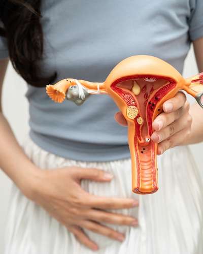
Endometriosis occurs when tissue similar to the lining of the uterus grows outside the uterine cavity. When this tissue involves the bladder, it is called bladder endometriosis.
The disease may be located on the outer surface of the bladder or may extend deeper into the bladder wall, particularly the detrusor muscle. Ultrasound studies describe bladder endometriosis as a nodule or thickening that may extend from the vesicouterine space into the bladder wall.
Bladder endometriosis can occur along with endometriosis involving other areas such as:
- Ovaries
- Uterosacral ligaments
- Pelvic peritoneum
- Bowel
- Ureters
- The area between the uterus and bladder
This is why evaluating only the bladder may not provide the complete picture. Endometriosis should be assessed as a disease that can involve multiple pelvic structures.
Symptoms vary from woman to woman, and some women may have relatively few urinary symptoms despite significant disease.
Common symptoms include:
- Pain while passing urine
Dysuria, or painful urination, can occur when endometriotic lesions involve the bladder.
A particularly important clue is when urinary discomfort becomes noticeably worse around menstruation.
- Blood in the urine during periods
Blood in the urine, medically called haematuria, particularly when it occurs cyclically around menstruation, can be an important warning sign.
However, haematuria has many possible causes, including urinary infections, stones and other urinary tract conditions. It should therefore always be medically evaluated rather than automatically attributed to endometriosis.
- Increased urinary frequency
Some women may feel the need to urinate more frequently or experience bladder irritation.
- Urgency
A sudden, difficult-to-control urge to urinate may occur in some patients.
- Lower abdominal or pelvic pain
Bladder endometriosis may cause pain in the lower abdomen, pelvis or behind the pubic bone.
- Painful periods
Severe dysmenorrhoea is one of the common symptoms associated with endometriosis. The presence of urinary symptoms that worsen during menstruation should increase suspicion of bladder involvement.
- Pain during intercourse
Deep pelvic endometriosis can cause deep dyspareunia, or pain during sexual intercourse. The presence of this symptom along with urinary complaints may suggest more extensive pelvic endometriosis.
- Difficulty conceiving
Endometriosis can also be associated with infertility. If a woman has infertility together with severe period pain, pelvic pain or cyclical urinary symptoms, endometriosis should be considered.
International endometriosis guidelines recognise symptoms such as dysmenorrhoea, deep dyspareunia, dysuria and haematuria among the clinical features that should prompt consideration of endometriosis.
One of the most useful clues is the relationship between symptoms and menstruation.
For example:
- Burning or pain while urinating mainly around periods
- Pelvic pain that becomes worse during menstruation
- Urinary urgency that increases around periods
- Blood in urine that appears cyclically
- Severe period pain associated with urinary complaints
These patterns do not prove bladder endometriosis, but they should prompt a detailed gynaecological evaluation.
Ultrasound is not simply about looking at the uterus and ovaries.
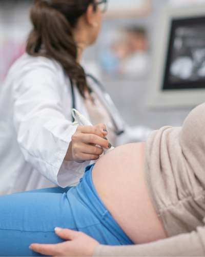
When endometriosis is suspected, a targeted pelvic ultrasound can be used to assess the pelvis systematically and look for signs of deep endometriosis.
Current guidelines recommend ultrasound in the diagnostic work-up of suspected endometriosis, including assessment for deep disease involving the bladder, bowel and ureters.
A specialist ultrasound can assess:
- The uterus and ovaries
- Ovarian endometriomas
- The bladder wall
- The space between the bladder and uterus
- Pelvic adhesions
- The mobility of pelvic organs
- Other areas suggestive of deep endometriosis
- Associated bowel or ureteric involvement
Specialist ultrasound can be particularly useful because the examination is not performed in isolation.
An ultrasound report may say that the uterus and ovaries appear normal. But this does not necessarily mean that endometriosis is absent.
Superficial endometriosis can be difficult to detect on imaging, and ultrasound accuracy depends considerably on the expertise of the person performing and interpreting the examination.
This is why clinical correlation is extremely important.
A gynaecologist considers:
- Your age
- Menstrual history
- Severity and duration of period pain
- Urinary symptoms
- Whether symptoms are cyclical
- Pain during intercourse
- Bowel symptoms
- Previous endometriosis diagnosis or surgery
- Infertility history
- Previous treatment and response
- Ultrasound findings
The combination of symptoms, examination and imaging can provide a much clearer picture than any one test alone.
In other words, an ultrasound should not simply be read as a list of measurements. It should be interpreted in the context of the patient’s symptoms and clinical history.
No.
A normal ultrasound does not completely exclude endometriosis, particularly superficial peritoneal disease.
However, a specialist ultrasound can be highly valuable for identifying deep endometriosis and planning treatment.
When deep endometriosis is suspected, specialist ultrasound or MRI may be recommended to determine the extent and location of disease.
This distinction is important:
Normal imaging does not always mean “no endometriosis.”
Instead, the clinical picture must be considered as a whole.
Knowing where the disease is located before surgery helps the surgical team plan the procedure.
Bladder endometriosis can sometimes extend into the bladder muscle and may be associated with disease around the ureters or other pelvic structures.
Preoperative assessment can help answer important questions:
- Where exactly is the lesion?
- How deeply does it extend?
- Is the bladder muscle involved?
- Is the lesion close to the ureteric openings?
- Is there associated bowel or pelvic endometriosis?
- Will bladder surgery be required?
- Is cystoscopy required before or during surgery?
Guidelines recommend specialist imaging for suspected deep endometriosis so that the extent of disease can be assessed and appropriate treatment planned.
Treatment depends on the severity of symptoms, location and depth of disease, reproductive plans and the presence of other endometriotic lesions.
Medical treatment may help control endometriosis-related pain and suppress disease activity in appropriate patients. However, when significant bladder endometriosis requires surgical treatment, complete excision of the lesion may be considered.
For deep bladder lesions involving the bladder muscle, partial bladder resection or excision of the affected segment may be required.
The goal is not simply to “burn” a visible spot. The surgeon needs to identify the extent of the lesion and remove the affected endometriotic tissue while preserving as much healthy bladder tissue as safely possible.
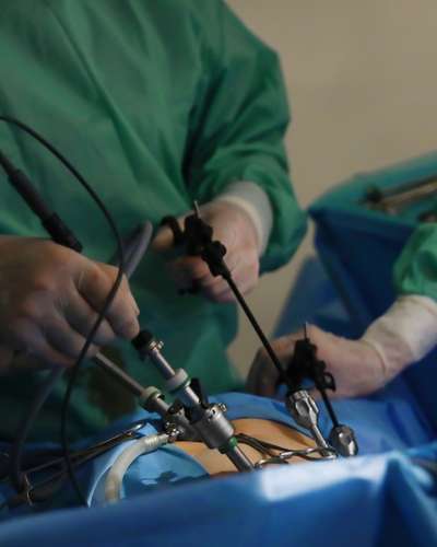
Laparoscopy allows the surgeon to examine the pelvis using magnification and small abdominal incisions.
For endometriosis surgery, this can provide several advantages:
- Magnified visualisation of pelvic anatomy
- Better identification of deep endometriotic lesions
- Precise dissection around important structures
- Treatment of associated pelvic endometriosis during the same procedure
- Smaller abdominal incisions
- Less postoperative wound discomfort compared with open surgery in appropriate cases
- Faster recovery for many patients
For bladder endometriosis specifically, surgical approaches can include laparoscopic excision or partial cystectomy depending on the depth and location of the lesion.
3D laparoscopy provides stereoscopic depth perception during minimally invasive surgery.
This can be particularly useful during complex endometriosis surgery because the pelvis contains closely positioned structures such as:
- Bladder
- Uterus
- Ureters
- Bowel
- Major blood vessels
- Pelvic nerves
Endometriosis can also cause dense adhesions and distortion of normal anatomy.
With 3D visualisation, the surgeon can better appreciate tissue planes and depth while performing delicate dissection.
However, it is important to understand that 3D laparoscopy itself does not automatically guarantee complete removal of endometriosis. The outcome depends on appropriate patient selection, detailed preoperative mapping, surgical expertise, careful identification of disease and safe excision.
For surgically treated bladder endometriosis, the objective is to remove the endometriotic nodule or affected tissue completely while protecting healthy bladder and surrounding structures.
Published surgical literature describes laparoscopic excision and partial cystectomy as approaches for bladder endometriosis, with cystoscopy helping to identify the lesion and its relationship to the ureteric openings.
A stepwise approach may include cystoscopy, assessment of the pelvis, mobilisation of the bladder, excision or partial cystectomy when required, bladder closure and a water-leak test.
In selected cases, combining laparoscopy with cystoscopy can help precisely locate the bladder lesion and guide complete excision while avoiding unnecessary removal of normal bladder tissue.
Endometriosis is often a multifocal disease.
A patient may have a bladder lesion together with endometriosis involving the ovaries, bowel, uterosacral ligaments or other pelvic structures.
Therefore, a comprehensive endometriosis surgery should evaluate the entire pelvis rather than focusing only on the most obvious lesion.
This is another reason why detailed preoperative mapping and experienced endometriosis surgery are important.
You should consider a gynaecological evaluation if you experience:
- Severe or worsening period pain
- Pain while passing urine, especially around periods
- Recurrent urinary symptoms without a clear explanation
- Blood in urine, particularly if it appears cyclically
- Chronic pelvic pain
- Deep pain during intercourse
- Bowel symptoms that worsen during periods
- Difficulty conceiving along with symptoms suggestive of endometriosis
- Previous endometriosis surgery with recurrent symptoms
Blood in the urine should always be evaluated medically because urinary tract infections, stones and other conditions can cause haematuria as well.
Bladder endometriosis can be challenging because the symptoms may initially look like a urinary problem.
The key is to connect the symptoms with the menstrual cycle and evaluate the pelvis systematically.
A gynaecologist-led ultrasound, interpreted alongside the clinical history, can help identify deep endometriosis and guide further evaluation. In suspected deep bladder disease, specialist ultrasound or MRI may be used to define the extent of involvement.
When surgery is required, minimally invasive excision using laparoscopy, with cystoscopic assessment when appropriate, can allow the surgeon to precisely identify and remove bladder endometriotic lesions while preserving healthy tissue.
For complex cases, 3D laparoscopy can provide enhanced depth perception and visualisation during delicate pelvic dissection.
If you are experiencing severe period pain, cyclical urinary symptoms, pelvic pain or other symptoms suggestive of endometriosis, an expert gynaecological evaluation can help identify the underlying cause.
Dr Uma Mishra at Gayatri Hospital, Raipur provides gynaecological care with a focus on minimally invasive laparoscopic management of endometriosis.
Gayatri Hospital is located in Rohinipuram, Raipur, Chhattisgarh.
If bladder endometriosis is suspected, appropriate evaluation may include detailed clinical assessment, specialist pelvic ultrasound and, where required, MRI, cystoscopy and minimally invasive surgical management.
The goal is not merely to treat the symptoms, but to identify the extent of endometriosis and develop an appropriate treatment plan for the individual patient.
Bladder involvement is uncommon compared with pelvic endometriosis overall, but it is the most common form of urinary tract endometriosis.
No. Haematuria can occur, particularly when symptoms are cyclical, but many patients may have pelvic pain, dysuria, frequency or other symptoms without visible blood in the urine.
Yes. Specialist transvaginal ultrasound can identify many cases of deep bladder endometriosis and is an important part of the diagnostic work-up. However, a negative ultrasound does not completely exclude all forms of endometriosis.
No. The need for MRI depends on the clinical situation and ultrasound findings. MRI can be particularly useful for mapping the extent of deep disease and its relationship with structures such as the ureters or when complex disease is suspected.
In appropriately selected patients, yes. Laparoscopic excision or partial bladder resection can be used depending on the depth and location of the disease. Cystoscopy may be performed before or during surgery to help localise the lesion.
3D laparoscopy provides stereoscopic depth perception and may be particularly useful for complex pelvic dissection. However, successful endometriosis surgery depends on the surgeon’s experience, disease mapping, surgical technique and appropriate case selection—not simply on the equipment used.
Endometriosis can recur after treatment. The risk depends on factors including the extent of disease, completeness of excision, hormonal environment and other individual factors. Long-term follow-up may therefore be appropriate, particularly in patients with deep endometriosis involving the bladder or ureter.
Dr. Uma Mishra
Consultant Laparoscopic Gynecologist, Raipur
Specializing in advanced minimally invasive gynecological procedures and high-risk obstetric care, Dr. Mishra has extensive experience in managing complex pregnancy cases with precision and compassion.

Precision. Compassion. Women’s Health Redefined.
Years’ Experience
0
+
Laparoscopic Surgeries
0
+
Comprehensive health care for women of all age group
Advanced Laparoscopy
Minimally invasive, highly precise & advanced surgical treatments
Gynecologic Treatments
All issues related to periods, pelvic pain, hormonal imbalance & post menopause.
Pregnancy
Care
From cesarean section to painless delivery and expert care for high risk pregnancy.

Infertility Treatments
Accurate diagnosis and advanced treatment options for infertility issues.
Learn about bladder endometriosis symptoms, diagnosis, specialist ultrasound and minimally invasive 3D laparoscopic treatment in Raipur with Dr Uma Mishra.
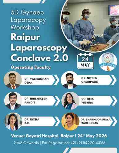
Join Raipur Laparoscopy Conclave 2.0 on 24 May 2026 at Gayatri Hospital, Raipur. Learn advanced 3D gynec laparoscopic techniques through live surgeries including TLH, myomectomy, endometriosis surgery, radical hysterectomy, VVF repair, and more from expert faculty across India.

Experience advanced 3D laparoscopy in Raipur for fibroids, endometriosis & gynecologic conditions. Dr Uma Mishra, Endometriosis & gynec cancer expert surgeon in Raipur.

Learn about laparoscopic transabdominal cerclage in Raipur by Dr. Uma Mishra. A highly effective solution for cervical incompetence and recurrent pregnancy loss.